
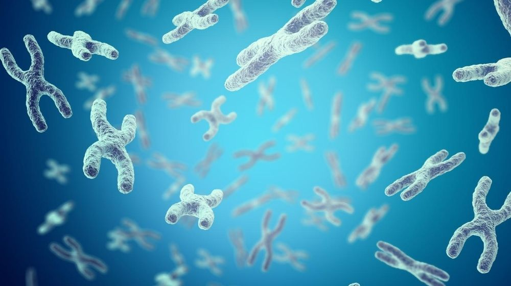
At the end of every strand of DNA inside a cell sits a specialized sequence of protective cap structure. Think of them like the plastic aglets on the end of shoelaces, preventing the genetic code from fraying or fusing with neighboring chromosomes during division. These caps are telomeres. Every single time a human somatic cell divides, its machinery fails to replicate the very end of the linear DNA sequence. This is known as the end-replication problem. Bit by bit, with every cycle of division, these protective boundaries erode.
Cellular aging is governed directly by this progressive erosion. When telomeres become critically short, the cell recognizes the exposed chromosome ends as double-strand DNA breaks. Division stops cold. The cell enters a state called cellular senescence, or it undergoes programmed cell death. This finite limit on division; first identified by Leonard Hayflick in 1961; sets the clock for replicative aging across human tissues.
The Molecular Engine Behind Telomere Shortening
Why do these chromosome ends degrade so predictably? The standard DNA polymerase enzyme complex operates in a single direction, requiring an RNA primer to initiate synthesis on the lagging strand. Once that primer drops off, a small gap remains at the extreme tip of the chromosome. Human telomeres consist of thousands of repeats of a specific hexanucleotide sequence, TTAGGG, bound tightly by a six-protein complex called shelterin.
Shelterin acts as a physical shield. It prevents the cell’s innate DNA damage repair pathways from mistakenly welding chromosome end protection structures together. As telomere shortening strips away these repeats, shelterin disassembles. The cell senses an open wound. At that threshold, p53 and p21 signaling pathways activate, halting the cell cycle permanently.
It is not just basic replication that degrades these caps; environmental hazards inside the cell speed up the damage. Oxidative stress and DNA damage act as accelerants. Guanine bases, which make up half of the TTAGGG repeat, are exceptionally vulnerable to reactive oxygen species. A 2014 study published in Nucleic Acids Research demonstrated that oxidative DNA lesions within telomeric repeats are repaired far less efficiently than elsewhere in the genome. The physical structure of the telomere seems to trap oxidative damage, causing sudden, premature structural loss long before the end-replication problem would normally trigger senescence.
Countering Erosion with Enzymatic Repair
Biology does possess a countermeasure. The telomerase enzyme is a ribonucleoprotein complex capable of adding TTAGGG repeats back onto chromosome tips from scratch. It carries its own RNA template and a catalytic reverse transcriptase component. In healthy adult human biology, telomerase activity is tightly restricted; expressed almost exclusively in germline cells, certain stem cell niches, and developing embryonic tissue. Most somatic tissues carry virtually zero active telomerase.
Research into longevity research focuses heavily on whether transiently reactivating this enzyme can delay biological aging without raising the risk of unchecked cell growth. Because cancer cells rely on telomerase activation to achieve immortality, any intervention targeting this pathway requires delicate balance.
Laboratory models demonstrate that restoring telomerase can reverse specific markers of tissue degeneration. Mice engineered with telomerase deficiencies show premature tissue atrophy, immune decline, and shortened lifespans; defects that clear up when telomerase gene expression is restored. Translating these findings to controlled human models remains one of the primary goals in cell biology.
Investigators studying chromosomal maintenance frequently rely on standardized biochemical tools to probe cellular senescence in vitro. Synthetic research peptides serve as vital tools in these laboratory environments, allowing researchers to observe how specific amino acid sequences interact with cell signaling pathways. By isolated exposure to research peptides, molecular biologists can examine cell culture survival, peptide stability, and enzymatic expression under controlled stressors. High-purity compounds like wholesale research peptides from Elivena provide laboratories with consistent reagents needed to measure changes in telomerase expression, DNA repair markers, and cellular stress responses. The precise purity of research peptides ensures that observed changes in cell cultures reflect true biochemical reactions rather than experimental noise.
Measuring the Clock: Telomeres and Epigenetic Clocks
Measuring biological age requires clear metrics, but telomere length alone tells only part of the story. While telomere length tracks cell division history, newer molecular tools look at different layers of cellular data. Epigenetic clocks monitor specific DNA methylation patterns across the genome, offering a distinct measurement of biological aging that operates parallel to chromosome cap loss.
Data shows that telomere attrition and epigenetic modifications track slightly different aspects of the aging process. A person can present with relatively preserved telomeres while displaying an advanced epigenetic age due to metabolic stress or chronic inflammation. Combining both measurements yields a significantly clearer picture of cellular health.
Researchers often categorize the biological drivers of cellular decline into distinct, intersecting mechanisms:
- Telomere exhaustion and shelterin destabilization limiting division limits
- Accumulation of senescent cells secreting pro-inflammatory signaling molecules
- Mitochondrial dysfunction driving systemic oxidative stress and DNA damage
- Epigenetic drift altering gene expression profiles over time
Targeting any single mechanism rarely stops the complex cascade of tissue aging; these systems push on one another continuously.
Interventions, Caloric Limits, and Senolytics
Can external factors change the rate of telomere loss? Extensive observational studies link lifestyle factors and telomere length closely. High physical activity, low systemic inflammation, and minimal exposure to chronic psychological stress correlate consistently with longer telomeres in white blood cells.
Caloric restriction studies provide some of the most consistent laboratory data regarding longevity research. In animal models, reducing caloric intake without malnutrition reduces systemic oxidative stress, downregulates nutrient-sensing pathways like mTOR, and preserves telomere length over extended periods. Less metabolic waste means fewer reactive oxygen species damaging guanine-rich telomeric DNA.
Another rising focus is the study of senolytics; molecules designed to selectively destroy senescent cells that have already reached their division limit. Senescent cells refuse to undergo normal cell death; instead, they secrete a cocktail of inflammatory cytokines that damage surrounding healthy cells and accelerate neighboring telomere loss. Clearing these stubborn, dysfunctional cells in rodent models reduces tissue inflammation and improves functional markers across multiple organ systems.
What Research Shows About the Limits of Extension
Reversing cellular aging requires more than keeping telomeres long. A cell with artificially elongated telomeres can still accumulate damaged proteins, suffer mitochondrial failure, or develop lethal mutations elsewhere in its genetic code.
Experiments in animal models reveal that simply overexpressing telomerase can increase the incidence of tumor formation if total genomic stability is not maintained. Cancer cells hijack telomerase to achieve infinite division capacity; roughly 85 to 90 percent of human malignant tumors express high levels of the enzyme. Safe biological interventions must find a way to maintain chromosome end protection without giving damaged cells free rein to divide indefinitely.
Slowing down cellular aging remains a puzzle with multiple interlocking pieces. Telomere shortening is not the sole cause of aging, but it acts as a primary counting mechanism for cellular life spans. Telomeres set a hard limit on replication that prevents damaged, mutated cells from continuing to propagate. Unlocking the ability to tune this system; preserving healthy tissue function while maintaining tumor suppression; represents one of the most promising avenues in modern molecular biology. Laboratory work across cell culture models, senolytic therapies, and biochemical research peptides continues to push closer toward understanding how these cellular clocks can be safely adjusted.